![]()
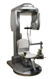
One of the things that sets our office apart from the crowd is our 3D imaging system.
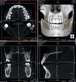
We routinely use our Prexion 3D Cone Beam Computed Tomography (CBCT) system in the assessment and planning for the placement of dental implants. In 2012, the American Academy of Oral and Maxillofacial Radiology (AAOMR) recommended that cross-sectional imaging be used for the assessment of all dental implants sites and stated that CBCT is the imaging method of choice for gaining this information. Our use of the Prexion 3D CBCT fully conforms with these recommendations.
What is CBCT?
Cone Beam Computed Tomography (CBCT) is a imaging technique that is used to generate a 3 dimensional image. The technology was first introduced into the United States in 2001 and has become increasingly important in the treatment planning involving dental implants. CBCT scans are fast and comfortable for our patients as nothing is even inserted into the mouth.
In just 17 seconds, our Prexion 3D system will acquire 512 individual views that are reconstructed into a 3D image of the patient's jaw anatomy. This image can be used to access the height, width, and density of the bone at potential dental implant placement sites. This information is important to ensure that implants are placed in the safest possible manner with the highest possible success rates.
The Prexion 3D system can also be used for many other uses such as:
- Evaluating the proximity of nerves to impacted wisdom teeth
- Evaluating pathology located in the jaw
- Evaluating possible root fractures